
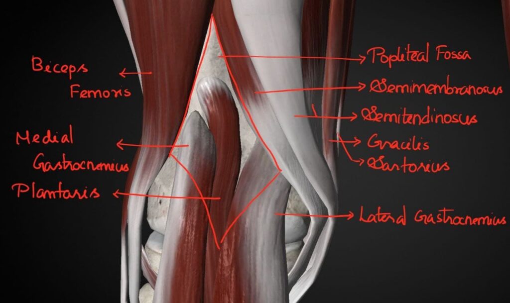
Popliteal fossa
Introduction:
- Popliteal fossa is a shallow diamond shaped fossa at the back of the knee.
- This corresponds to the triangular shaped Cubital fossa in the forearm.
- Most importantly it is felt best during the Semi-flexed position of the knee joint.
Location:
- It is a diamond-shaped depression located behind the knee joint.
- Moreover behind the lower part of the Femur and upper part of the Tibia.
Boundaries of Popliteal Fossa:
Superolaterally :
- Biceps femoris.
Superomedially:
- Semitendinosus and the Semimembranosus.
Moreover Gracilis, Sartorius and Adductor magnus supplements the superomedial boundary.
Inferolaterally :
- Lateral head of Gastrocnemius.
Moreover Planatris also bounds the fossa inferolaterally.
Inferomedially:
- Medial head of Gastrocnemius.
Roof:
Roof of the fossa is formed by Deep Fascia/Popliteal fossa.
The superficial fascia over the roof contains the following structures.They are:-
- Small saphenous vein and cutaneous nerves.
- Three cutaneous nerves.They are,
- Firstly Terminal part of posterior cutaneous nerve of thigh
- Secondly Posterior division of the medial cutaneous nerve of thigh.
- Finally the Peroneal or sural communicating nerve.
Floor:
From above downwards,
- Firstly, Popliteal surface of Femur.
- Secondly, Capsule of the Knee joint and the oblique popliteal ligament.
- Finally, Popliteal fascia covering the Popliteus.
Contents of the Popliteal fossa:
- Firstly- Popliteal artery, Popliteal veins and their branches and tributaries respectively.
- Secondly-Tibial nerve and its branches.
- Most importanly, Common peroneal nerve and its branches.
- The posterior cutaneous nerve of thigh.
- Genicular branch of the Obturator nerve.
- The Popliteal lymph nodes.
- Finally-Fat.
- The popliteal vessels and the tibial nerve are arranged one over the other.
- Structures arranged from deep to superficial are
- Firstly, Popliteal .
- Secondly, Popliteal vein.
- Finally, Tibial nerve.
Relations of the structures in the Popliteal fossa:
Upper part:
From medial to lateral side: Artery, Vein, Nerve.
Middle part:
From behind forwards: Nerve, Vein, and Artery.
Lower part:
From medial to lateral side: Nerve, Vein and Artery.
Responses